
Observation under the inverted microscope
單元大綱
-
-
Allow the phytoplankton to stand in the glass slides until it precipitates (about 10 minutes), then observe it under an inverted microscope at about 200-400x (Figs. 9 and 10).
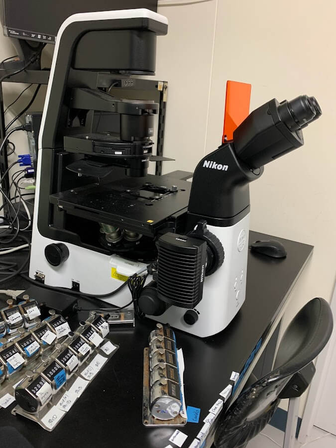
Fig. 9 Inverted microscope
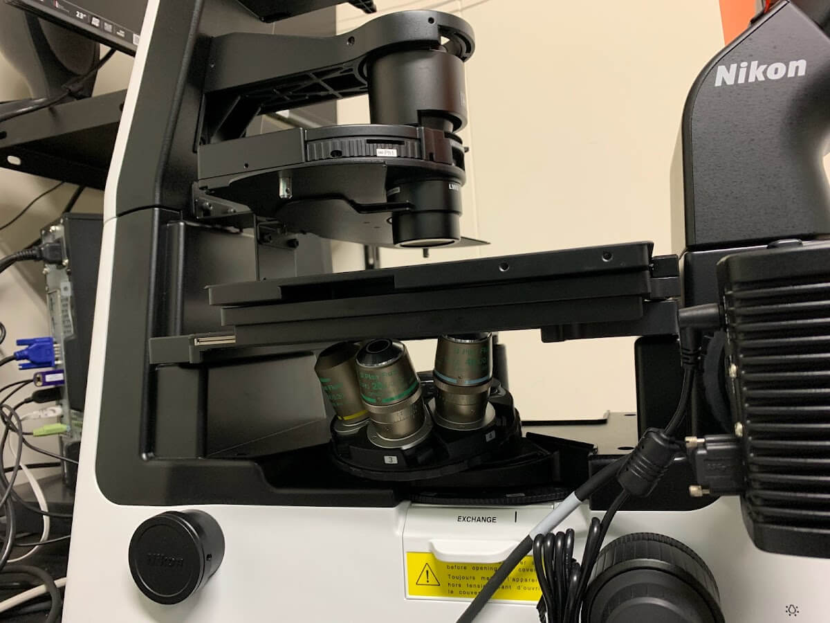
Fig. 10 Objective lens of an inverted microscope
-
During counting, the value of how many cells are present when counting the mesh is taken, and the number of observed cells per overall basal area is used to calculate how many cells were contained in the entire concentrated sample collected (Fig. 11).

Fig. 11 Counting method
*Count the cells “inside” the frame.
Even if there are no cells inside the compartment, the number of compartments is counted as 1.
For phytoplankton, identification is made to the species level based on Hasle and Syvertsen (1997) and Horner (2002). For the dinoflagellate Karenia selliformis, the number of chloroplasts in the cell is useful for species identification (Iwataki et al. 2021). For ciliates, the counts are divided into two groups: tintinnids and oligotriches. Nanoplankton and siliceous flagellates are also counted. A minimum of 300 cells per sample will be counted. After counting, the cell density (cells mL-1) of each taxon in the sample is calculated.
-