Phytoplankton observation of red tide in east Hokkaido
セクションアウトライン
-
-
After the fixed samples are weighed, they are placed on a stone table (Figs. 1 and 2) or other non-shaking surface for at least 24 hours to allow the phytoplankton to settle (Fig. 3).
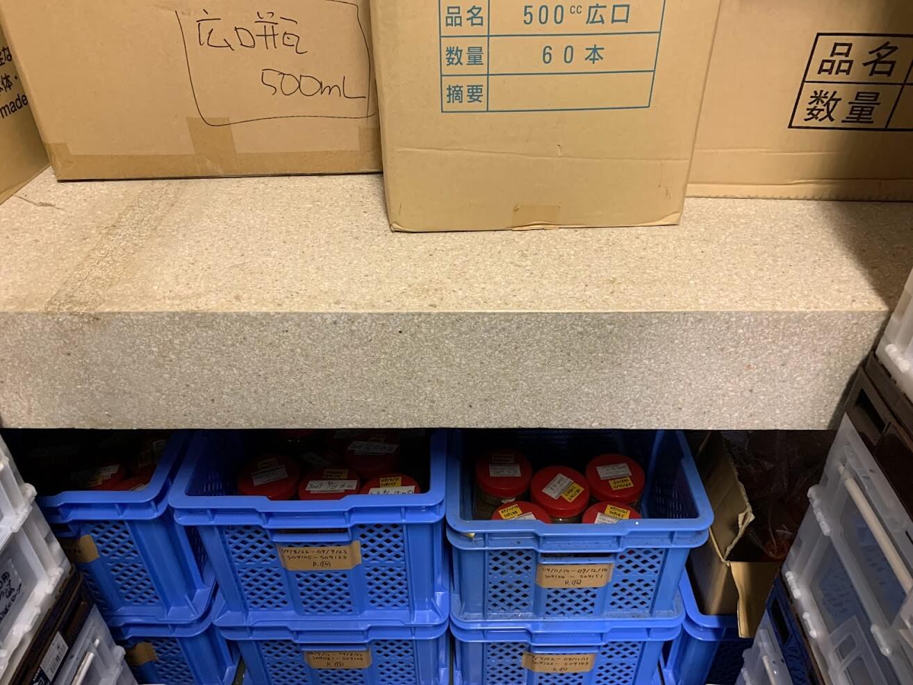
Fig. 1 Stone table
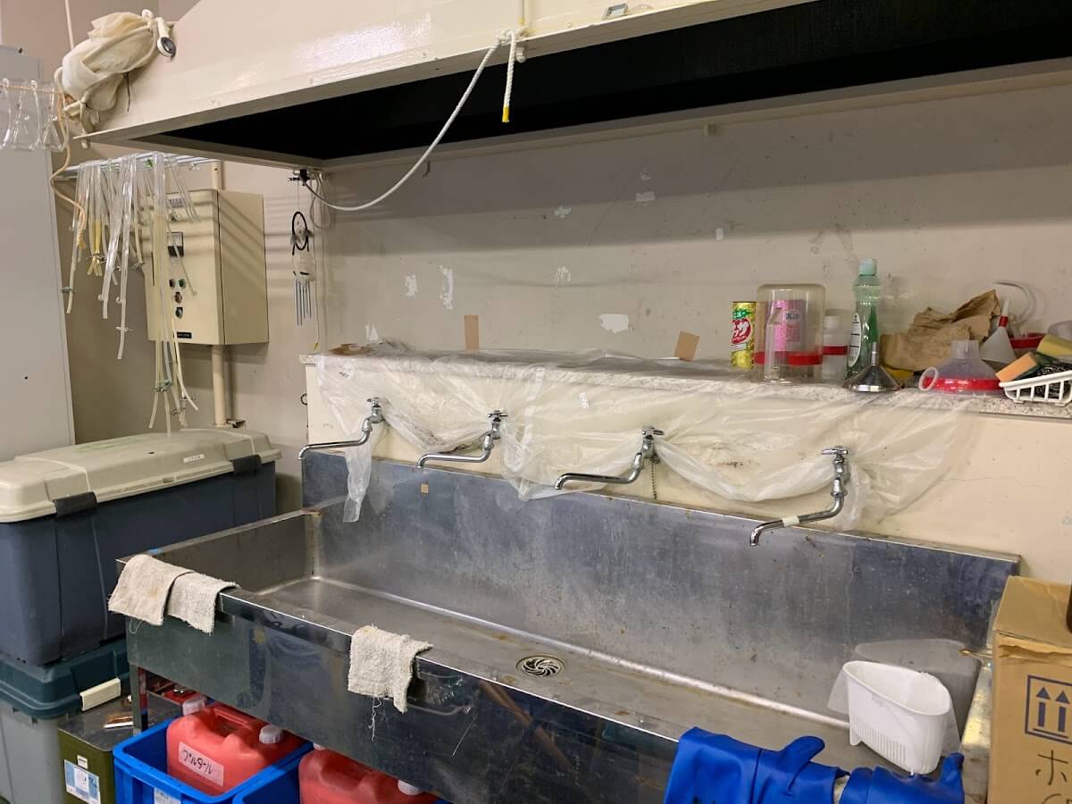
Fig. 2 Stone table
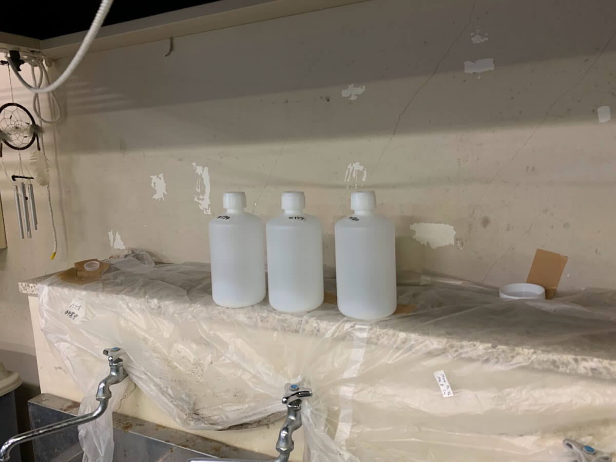
Fig. 3 Static precipitation on stone table
-
Then, the supernatant phytoplankton-free seawater is removed using a siphon (Fig. 4).
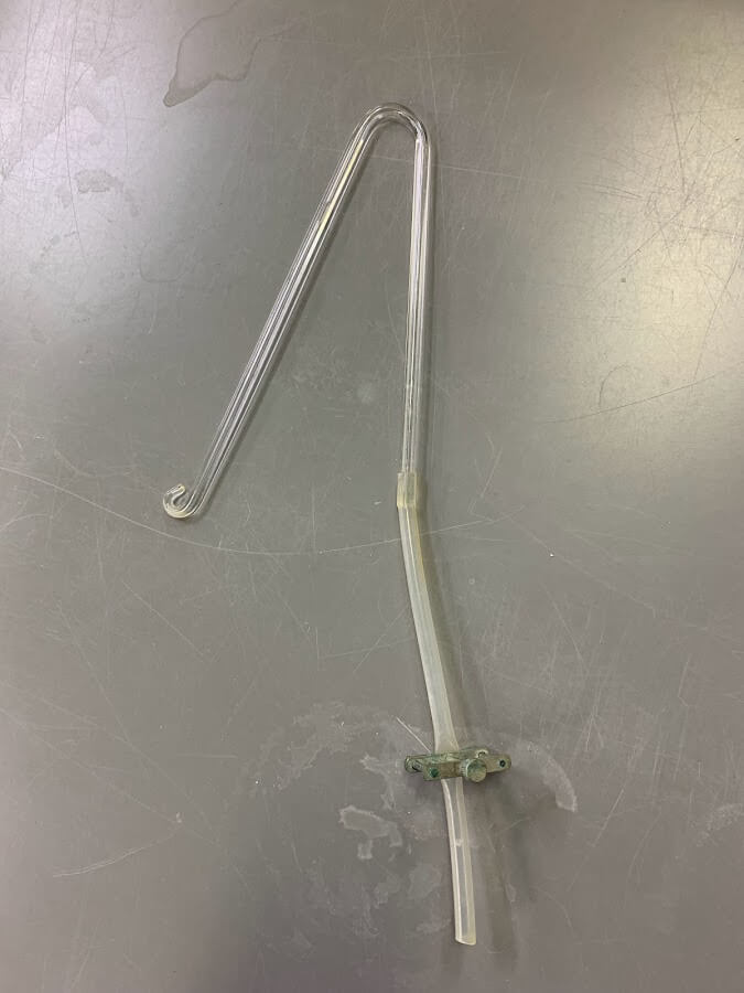
Fig. 4 Siphon
-
In the Plankton Laboratory, after concentrating in a 1 L container, it is placed in a 110 mL bottle, and precipitation again. After that, the supernatant is removed using a siphon. Samples are concentrated until the final concentration is in a 20 mL bottle (Fig. 5).
The weight of the concentrated sample is also measured.
The value that subtracted respectively the weight of the bottle used in the first field and the weight of the bottle used at final is used to determine how many times the sample was concentrated and the concentration factor of the sample.
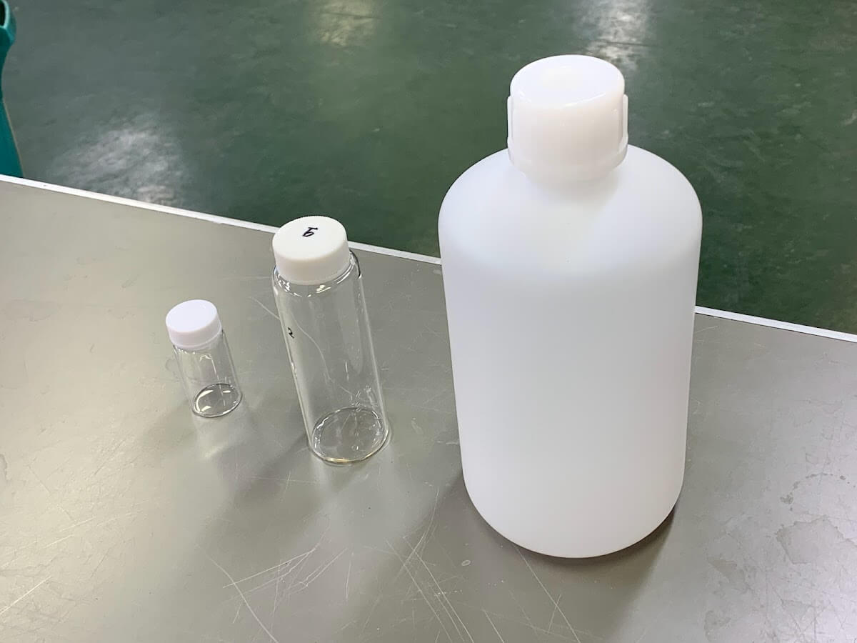
Fig. 5 Bottles used for concentration
-
-
-
The concentrated sample is also stored in the refrigerator to shield from light. At the time of samples microscopic examination, the sample is gently stirred and a fixed volume (about 2 mL) is collected with a pipette. It is placed on a glass slide for phytoplankton counting, which has a mesh pattern on the bottom (Fig. 6).
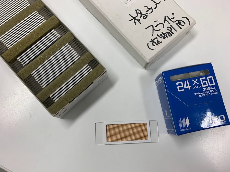
Fig. 6 Glass slides for counting
-
The glass slides for phytoplankton counting have a frame that can hold a 2 mL sample, into which the concentrated sample is pipetted.
A cover glass that fits this large frame is also commercially available.
When the sample is inserted between the glass slide frame and the cover glass, and the cover glass begins to float, capillary action causes the cover glass to rotate so that it fits over the glass slide frame (Figs. 7 and 8).
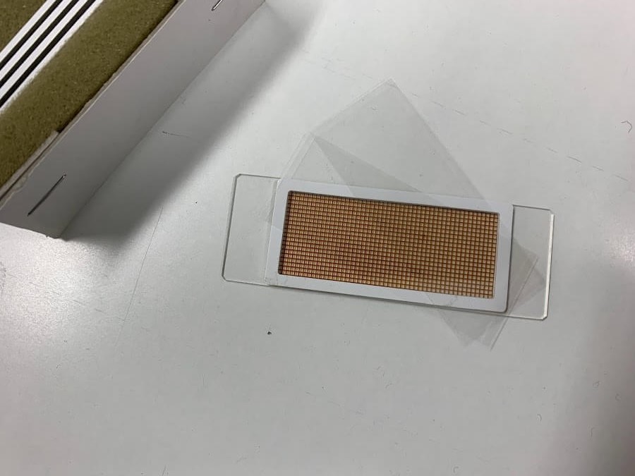
Fig. 7. Placing a cover glass on a counting glass slide
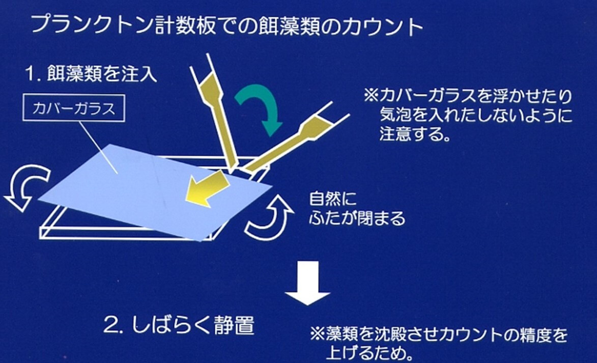
Fig. 8. Placing a cover glass
Counting algae on a plankton counting plate
1. Injecting algae
*Take care not to float the cover glass or introduce air bubbles.
The cover will close naturally.
2. Allow to stand for a while
*To allow algae to settle and improve counting accuracy.
-
-
-
Allow the phytoplankton to stand in the glass slides until it precipitates (about 10 minutes), then observe it under an inverted microscope at about 200-400x (Figs. 9 and 10).
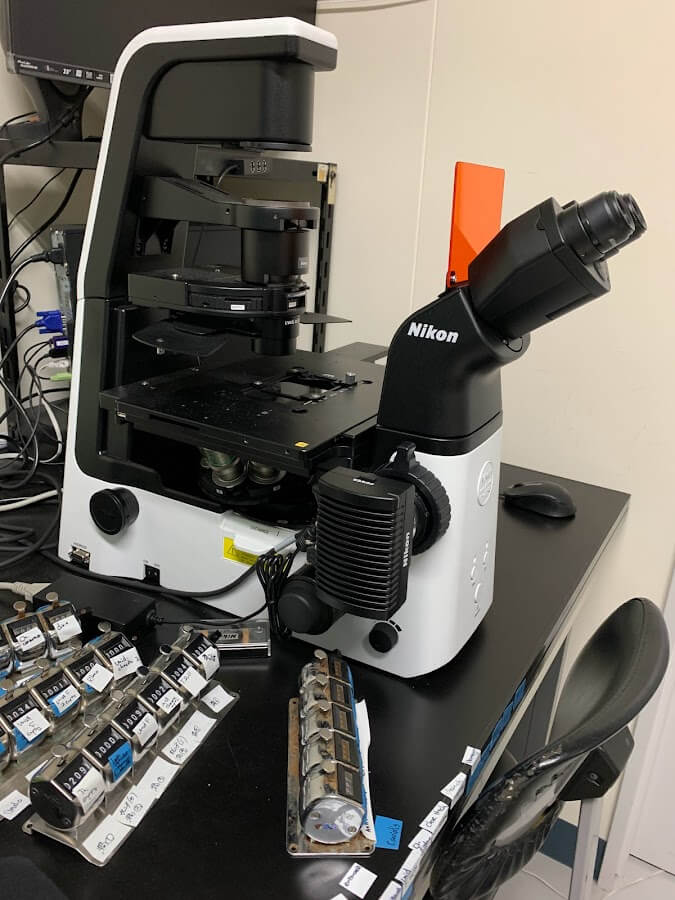
Fig. 9 Inverted microscope
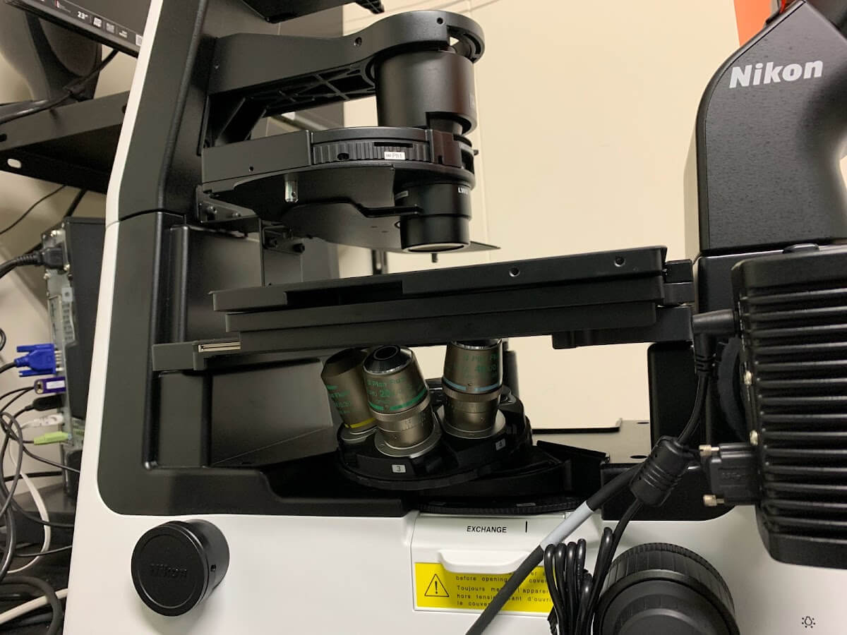
Fig. 10 Objective lens of an inverted microscope
-
During counting, the value of how many cells are present when counting the mesh is taken, and the number of observed cells per overall basal area is used to calculate how many cells were contained in the entire concentrated sample collected (Fig. 11).
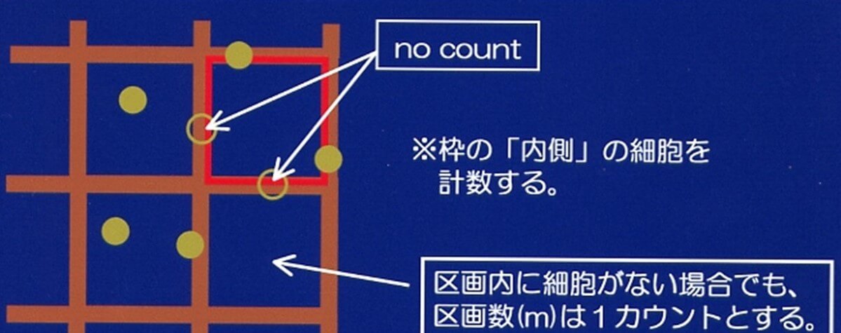
Fig. 11 Counting method
*Count the cells “inside” the frame.
Even if there are no cells inside the compartment, the number of compartments is counted as 1.
For phytoplankton, identification is made to the species level based on Hasle and Syvertsen (1997) and Horner (2002). For the dinoflagellate Karenia selliformis, the number of chloroplasts in the cell is useful for species identification (Iwataki et al. 2021). For ciliates, the counts are divided into two groups: tintinnids and oligotriches. Nanoplankton and siliceous flagellates are also counted. A minimum of 300 cells per sample will be counted. After counting, the cell density (cells mL-1) of each taxon in the sample is calculated.
-