We have developed a technology
to coat cell culture dishes with the collagen fibrils of sturgeons swim bladders.
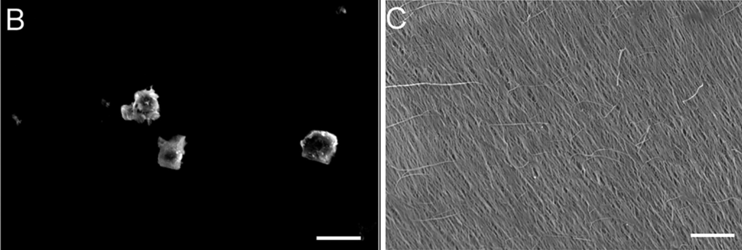
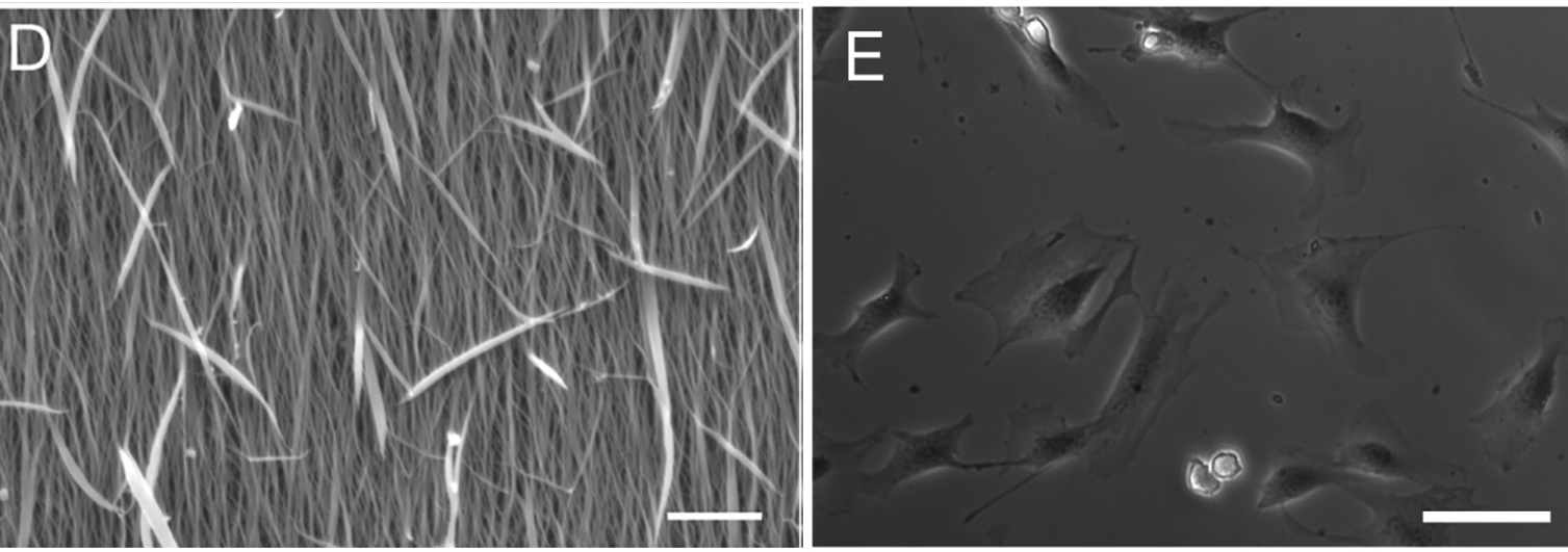
Scanning electron micrographs of a culture dish coated with (B) collagen
molecules,(C) thin fibrils,and (D) thick fibrils
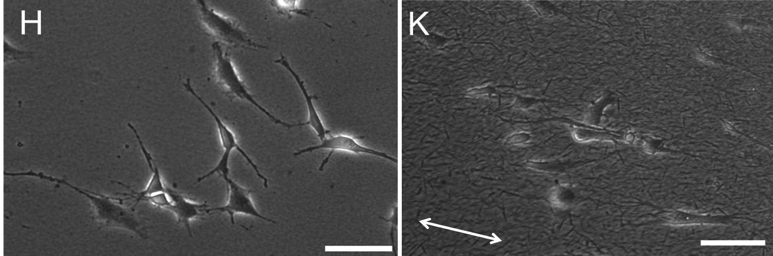
E, H, and K are polarized light micrographs of mouse osteoblast
progenitor cells cultured in each dish. Each shows a characteristic cell
morphology, and the cells, especially in K, extend in one direction along the
traveling direction of the fibrils (arrow in K). Although not shown in the
figure, cell proliferation and differentiation also differ greatly depending on
the type of coating. Such coating is not possible with porcine collagen. This
technology has made it possible to develop inventive research, such as investigating
the reaction of cells to collagen in detail.
We will further develop this technology, aiming to synthesize
three-dimensional cell scaffold materials in the future.
