Section outline

    • In the fertilized egg of a fish, the number of cells increases exponentially, leading to the formation of a blastocyst with approximately 1,000 cells. At this stage, the fate of the cells is not determined. Therefore, the development proceeds normally even if the cells of the embryo are removed or cells are transplanted from another embryo (Figure 1. Embryos obtained by cutting and replacing the blastocyst of a goldfish. Figure 2. Embryos in which the blastodisc cells are added or removed.


    • Figure 1Blastodisc transplantation at the blastocyst stage. A) Red-stained and unstained transparent embryos. B) Cutting the upper side of stained and unstained blastocysts. C) Immediately after cutting. D) Immediately after replacing the cut part and transplanting it. E) Restored blastodisc.


    • Figure 2Embryos where the entire blastodisc is transplanted to the animal pole side of another embryo using transplantation similar to Figure 1. Bottom (from left): transplanted embryo, embryo with the upper part of blastodisc removed, and untreated embryo. All develop normally.
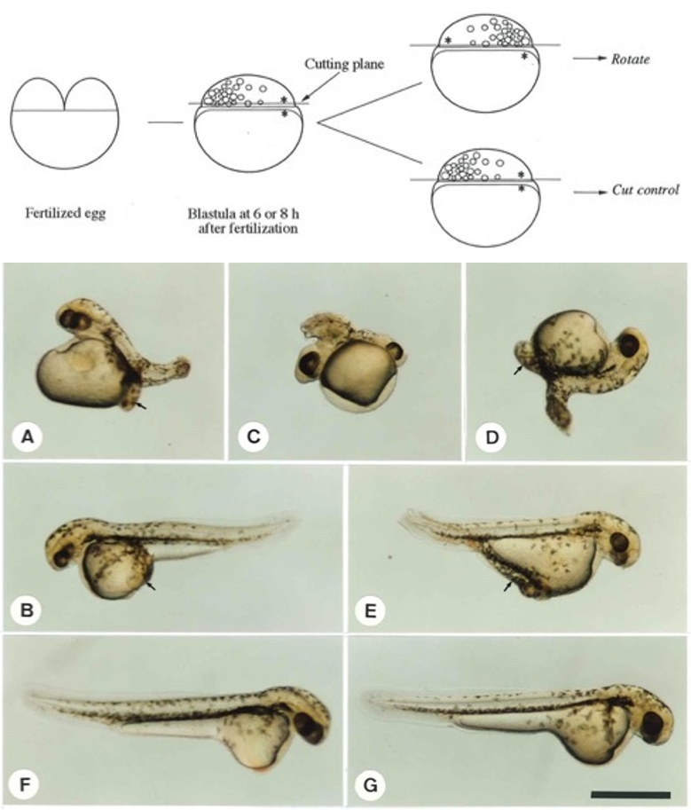
    • The direction of the differentiation of blastodisc cells in the blastocyst stage is determined by the induction signal from the yolk side. The yolk is also called the yolk cell because the nucleus also enters the yolk side during the division process of the fertilized egg. When the blastodisc of the anaphase blastocyst stage is separated from the yolk cell, rotated 180° horizontally and reattached to the yolk cell, many individuals with two bodies are born from this embryo. This is because the body is made up of two groups of cells that have started building the body before or after being separated from the yolk cell (Figure 3Individual with two axes generated from an embryo in which the blastodisc of the anaphase blastocyst of a goldfish is separated, rotated 180°, and reattached.)


    • Figure 3A–E) Embryos in which the blastodisc in the blastocyst stage was separated from the yolk cell and rotated 180° horizontally and then reattached. A–D were operated on during the metaphase blastocyst stage, while B and E were operated on in the anaphase blastocyst stage. F is an embryo that was rotated 360° and reattached, and G is an untreated embryo.

    • The body-inducing power of these yolk cells originates in the embryo immediately after fertilization. Immediately after fertilization, if the embryo is cut in half between the animal pole side with the nucleus and cytoplasm and the plant pole side with a lot of yolk, embryonic formation no longer occurs, although the cells increase and grow longer, accompanied by the production of some cells. Furthermore, if it is cut closer to the animal pole side, it only becomes a cell mass. This indicates that the factors that shape future individuals are localized in the isolated plant pole side of the egg immediately after fertilization.


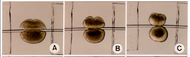
    • Figure 4. When a 2-cell stage embryo is pushed through with a silkworm gut, it is divided into the animal pole hemisphere with the cytoplasm and the plant pole hemisphere with a lot of yolk.
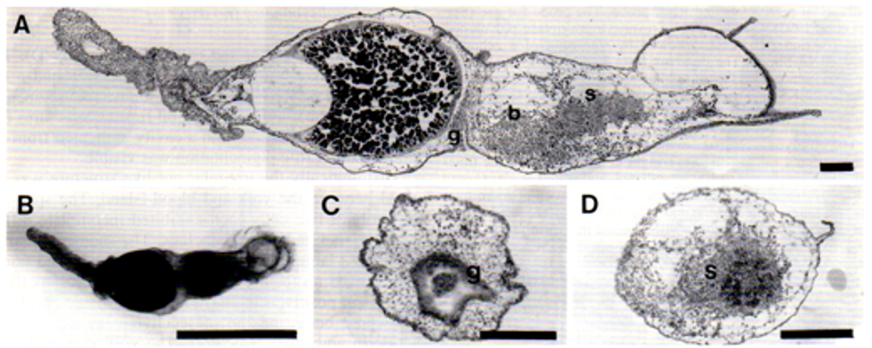

    • Figure 5When the animal pole hemisphere in the early cleavage stage is isolated and cultured, the embryo does not form a normal shape and it becomes an embryo with rotational symmetry.


    • Figure 6Removal of the cytoplasm at the 1-cell stage leads to the formation of an embryo with only a cell mass (K and L).